New microscope designed to reveal the function of individual genes in the brain
The new method could help to identify genes and their role in neuropsychiatric disorders
Changes in gene activity are fundamental to many neuropsychiatric disorders. However, the nature of the genes affected, the time and place in which these changes occur, or the impact of these changes on nerve cells, remain largely unknown. One of the reasons for this ignorance is the inability to tag individual genes and their products without disrupting normal cell functions. An international team from the Max Planck Institute of Neurobiology, the University of Birmingham, the Wigner Institute in Budapest and Femtonics Ltd. now aim to close this gap in methodology. The European Union supports the project with a Horizon 2020 grant, worth more than four million Euros.
Many diseases also have a genetic component. For example, gene mutations can lead to the development of cancer or psychiatric disorders such as schizophrenia, depression or autism. Most of these diseases seem to be caused not by a single mutation but are rather the result of the interplay of dozens, maybe even hundreds of altered gene functions. It would therefore be of great benefit for the investigation, diagnosis and therapy of these illnesses, to identify the affected genes and monitor their activity in the brain of the living organism.

An interdisciplinary team around Herwig Baier from the Max Planck Institute of Neurobiology, Attila Sik from the University of Birmingham, Miklós Veres from the Wigner Institute and the microscope developers at Femtonics Ltd. aim to develop such a method. Their plan is to tag the genetic building blocks in such a way that they can watch the activity and function of individual genes with the aid of a special microscope. These “live” observations should be possible during development as well as in later life.
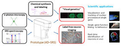
The new method, which will found a new field called “visual genetics”, combines techniques from two-photon microscopy, Stimulated Raman Spectroscopy (SRS) based on so-called Raman scattering, and the synthesis of specific marker molecules. The scientists’ aim is to visualize gene activity first in the brains of zebrafish and later on in the mouse brain. The project will be funded initially for three years.


